Recognizing the Symptoms of Retinal Detachment: A Guide to Early Detection
- September 4, 2024
- 0 Likes
- 269 Views
- 0 Comments
Introduction
Retinal detachment is a medical emergency that requires immediate attention to prevent permanent vision loss. This serious condition occurs when the retina, the light-sensitive layer of tissue at the back of the eye, becomes separated from its normal position. The retina is critical for vision because it captures light and sends visual signals to the brain. When it detaches, these signals are disrupted, and if left untreated, retinal detachment can lead to blindness. Understanding and recognizing the early symptoms of retinal detachment is crucial for prompt treatment and minimizing long-term damage.
What Is Retinal Detachment?
Retinal detachment occurs when the retina is pulled away from its normal position at the back of the eye. There are three main types of retinal detachment:
- Rhegmatogenous Retinal Detachment:
The most common form, this occurs when a tear or hole in the retina allows fluid to seep beneath it, causing the retina to lift away from the underlying tissue. - Tractional Retinal Detachment:
This type occurs when scar tissue on the surface of the retina contracts, pulling the retina away from the back of the eye. It is often associated with diabetic retinopathy. - Exudative Retinal Detachment:
In this form, fluid builds up beneath the retina without any tears or breaks, often due to inflammatory conditions or injury.
Early Symptoms of Retinal Detachment
Recognizing the symptoms of retinal detachment early can significantly improve the outcome of treatment. Some of the most common warning signs include:
- Sudden Onset of Floaters:
Floaters are tiny specks or strands that drift through your field of vision. While floaters can be common with age, a sudden increase in the number of floaters, especially when accompanied by other symptoms, may indicate retinal detachment. - Flashes of Light (Photopsia):
Flashes of light, which may appear as brief, bright bursts or flickers, are a key symptom of retinal detachment. These flashes often occur in the peripheral vision and may resemble seeing “stars” after being hit on the head. - A Shadow or Curtain Over Vision:
One of the more alarming symptoms of retinal detachment is the sensation of a dark shadow or curtain moving across your vision. This can start in one area of your peripheral vision and gradually spread as the detachment worsens. - Blurry or Distorted Vision:
As the retina detaches, vision may become blurry, distorted, or wavy. The severity of this symptom depends on the extent and location of the detachment. - Reduced Peripheral Vision:
Retinal detachment often affects the peripheral vision first. If you notice a loss of side vision or a narrowing of your visual field, this could be a warning sign.
Causes and Risk Factors for Retinal Detachment
Several factors can increase the risk of developing retinal detachment. These include:
- Aging: As we age, the vitreous gel in the eye becomes more liquid and can shrink, pulling on the retina and leading to tears or detachment.
- Previous Retinal Detachment: If you’ve had retinal detachment in one eye, you are at higher risk of it occurring in the other eye.
- Severe Nearsightedness (Myopia): Individuals with severe nearsightedness have longer eyeballs, which can cause the retina to become thinner and more prone to detachment.
- Eye Injuries: Trauma to the eye, such as from sports or accidents, can increase the risk of retinal detachment.
- Eye Surgery: Certain eye surgeries, particularly cataract removal, may increase the likelihood of retinal detachment.
- Family History: A family history of retinal detachment can raise the risk.
- Diabetes: Diabetic retinopathy, a complication of diabetes, can lead to scar tissue formation on the retina, resulting in tractional detachment.
Real-World Case Study
Case Study: David’s Sudden Vision Loss
David, a 55-year-old man with severe nearsightedness, experienced a sudden increase in eye floaters and flashes of light in his left eye. At first, he ignored the symptoms, attributing them to eye strain from long hours at work. However, two days later, he noticed a dark shadow creeping across his peripheral vision. Alarmed, David visited an ophthalmologist who diagnosed him with rhegmatogenous retinal detachment. He underwent surgery the next day to reattach the retina. Thanks to prompt treatment, David regained most of his vision and now receives regular check-ups to monitor his eye health.
Diagnosis of Retinal Detachment
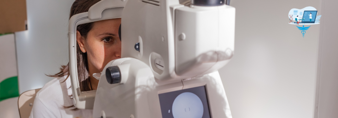
If retinal detachment is suspected, an ophthalmologist will perform a comprehensive eye exam, which may include:
- Dilated Eye Exam: Eye drops are used to widen the pupil, allowing the doctor to examine the retina and detect any tears, holes, or detachment.
- Ultrasound Imaging: If the retina is obscured by bleeding or other conditions, an ultrasound may be used to visualize the retina and confirm detachment.
- Optical Coherence Tomography (OCT): This non-invasive imaging test provides detailed cross-sectional images of the retina to assess its structure.
Treatment Options for Retinal Detachment
Treatment for retinal detachment usually involves surgery to reattach the retina and prevent further vision loss. The choice of surgery depends on the type and severity of the detachment.
- Laser Surgery (Photocoagulation):
In cases where there is a small retinal tear but no detachment, laser surgery can seal the tear and prevent fluid from passing through. This technique uses a laser to create small burns around the tear, causing scar tissue to form and seal the retina in place. - Cryopexy (Freezing Treatment):
Cryopexy involves freezing the area around the retinal tear to create scar tissue, which helps secure the retina to the back of the eye. This treatment is often used in conjunction with other surgical methods. - Pneumatic Retinopexy:
This procedure is used to treat smaller retinal detachments. A gas bubble is injected into the vitreous cavity of the eye, pushing the retina back into place. The patient is then instructed to maintain a specific head position to keep the bubble in place. Laser surgery or cryopexy is usually performed afterward to seal the tear. - Scleral Buckling:
In this procedure, a small silicone band (scleral buckle) is placed around the outside of the eye to gently push the wall of the eye against the retina. This helps relieve the pull on the retina, allowing it to reattach. - Vitrectomy:
For more complex or severe detachments, vitrectomy may be necessary. During this procedure, the vitreous gel is removed and replaced with a gas bubble or silicone oil to help reattach the retina. Over time, the gas is absorbed, or the oil may be surgically removed.
Prevention and Monitoring
While retinal detachment cannot always be prevented, regular eye exams can help detect early signs and reduce the risk of vision loss. Preventive steps include:
- Regular Eye Exams: Individuals with risk factors, such as severe nearsightedness or a family history of retinal detachment, should have regular eye check-ups to monitor retinal health.
- Protecting the Eyes from Injury: Wearing protective eyewear during activities that pose a risk of eye injury, such as sports, can help prevent trauma-induced detachment.
- Managing Underlying Conditions: For those with diabetes or other conditions that affect the eyes, managing these diseases can reduce the risk of complications like retinal detachment.
Conclusion
Retinal detachment is a serious condition that can lead to permanent vision loss if not treated promptly. Understanding and recognizing the early symptoms, such as flashes of light, sudden floaters, and shadowy vision, can be lifesaving. If you or someone you know experiences these symptoms, seek immediate medical attention. Timely intervention through surgical procedures can restore vision and prevent long-term damage.
For further information, visit:
- American Academy of Ophthalmology: https://www.aao.org
- Retina Society: https://www.retinasociety.org
- National Eye Institute: https://nei.nih.gov
References
American Academy of Ophthalmology. (2023). Retinal Detachment: Symptoms and Treatments. https://www.aao.org/eye-health/diseases/retinal-detachment
Retina Society. (2022). Understanding Retinal Detachment. https://www.retinasociety.org


Leave Your Comment